Medial Epicondyle Of Femur - How To Discuss
Medial Epicondyle Of Femur
What to do with the medial epicondyle of the femur
The medial epicondyle of the femur is a bony protrusion on the medial side of the distal end of the bone. Above the medial condyle has a height, the adductor tubercle, which serves to fix the superficial part or to insert the tendon of the adductor magnus.
So which ligament connects to the medial epicondyle of the femur?
medial collateral ligamentWhich muscle also attaches to the medial and lateral condyles of the femur?
Thigh muscleSecond, what muscles connect the medial epicondyle to the knee?
Structures on the medial side of the knee include tibia, femur, vastus medialis oblique, semitendinous tendon, gracilis tendon, Sartorius tendon, adductor-Magnus tendon, medial head of the gastrocnemius muscle, semitendinous tendon, membranous and medial meniscus, FL medial patellar femoral ligament (MPLM.
CL) eDoes the femur have epicondyles?
Epicondyle: The epicondyle provides sites for muscle attachment. Condyle: The medial and lateral condyles are examples of condyle. Epicondyle: The medial and lateral condyles of the humerus and femur are examples of the epicondyle. the function of each structure of the animal’s body.
What is the function of the medial collateral ligament?
The primary function of the medial collateral ligament is to prevent the leg from stretching too far inward, but it also helps keep the knee stable and rotatable. Damage to the medial collateral ligament usually occurs when the knee is struck directly from the outside.
What is the medial femoral condyle?
FMA. 32858. Anatomical bone terms. The medial condyle is one of the two protrusions on the lower end of the femur, the other being the lateral condyle. The medial condyle is larger than the lateral (external) condyle due to the increased weight load, since the center of gravity is median of the knee.
What is the lateral femoral condyle?
The lateral condyle is one of two processes in the lower part of the femur. The other is the medial condyle. The lateral condyle is the most prominent and is wider in the anterior and transverse diameters of the tobacco.
What is attached to the lateral femoral condyle?
Anatomical Bone Terms
Which Muscle Stabilizes the Knee in the Back?
What structure of the femur articulates with this patella surface?
Bone marrow stitches
Which ligament prevents the knee from moving backwards?
Posterior cruciate ligament
What does MCL thickening mean?
However, medial lateral lesions are heterogeneous. An important role for the stability of the ■■■■■ capsule, the medial side of the knee ■■■■■.
How can the growth of MCL be accelerated?
Hold crushed ice against the side of the injured knee for 15-20 minutes, repeating as needed, with 1 hour between treatments. Raising the knee in a chair or stool can help relieve discomfort. Protect the MCL as it heals to prevent further damage and reduce recovery time.
What happens to muscle and bone tissue when not in use?
How bad is MCL distortion?
Light In MCL sprains, the ligaments in the knee are slightly stretched, but not actually torn. You may have complaints of instability or a loose knee. In a severe Grade III MCL sprain, the ligament ruptures completely, causing swelling and sometimes bleeding under the skin.
Which muscle groups are most important for bending and extending the knee?
The quadriceps femoris muscle group (rectus femoris, vastus lateral, vastus medius, and vastus intermediate) crosses the knee through the patella and works to straighten the leg. The hamstring muscles (semitendinosus, semimembranous and hamstring) flex the knee and straighten the hip.
What is between the femur and the tibia?
In humans and other primates, the knee connects the thigh to the bone and consists of two joints: one between the femur and tibia (tibiofemoral ■■■■■) and one between the femur and patella (patellofemoral ■■■■■). It is the largest ■■■■■ in the human body.
Which ligament prevents valgus forces in the knee?
The medial collateral ligament (MCL) prevents lateral movement of the tibia over the femur when valgus is applied (away from the midline) to the knee. It passes between the medial epicondyle of the femur and the anteromedial aspect of the tibia.
Where is the medial side of the knee?
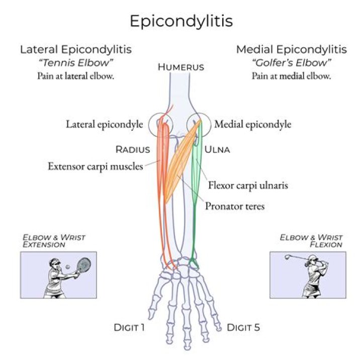
Which nerve breaks around the medial humeral condyle?
The ulnar nerve is medial to the median nerve as it passes through the arm. In the lower arm, it protrudes through the intermuscular septum and then goes posterior to the medial epicondyle of the humerus.